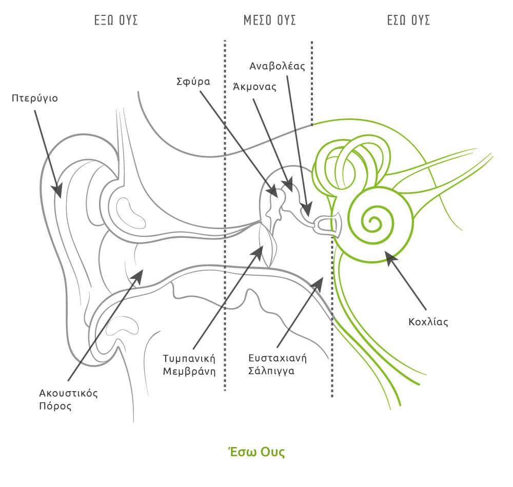
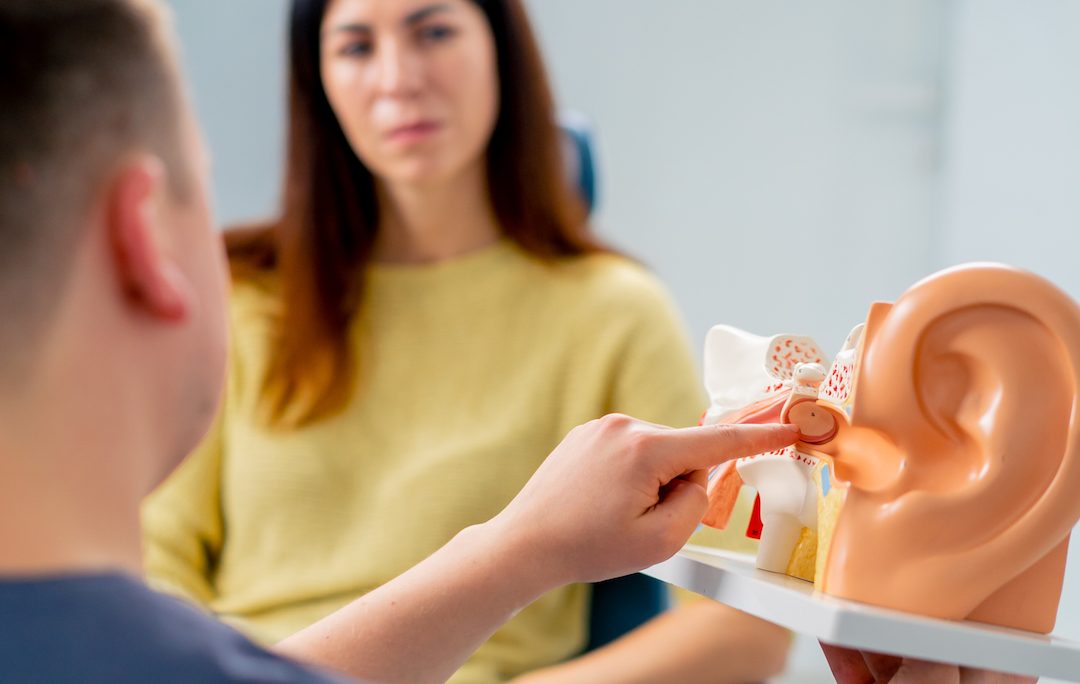
Βασική ανατομία και λειτουργίες του αυτιού
Έξω ους
Το έξω ους περιλαμβάνει το πτερύγιο, το οποίο μας βοηθά να αντιληφθούμε αν ο ήχος έρχεται από μπροστά ή από πίσω. Η παρουσία μάλιστα του πτερυγίου αυξάνει κατά 5dΒ την πίεση που ασκείται στο τύμπανό μας για τους ήχους που έρχονται από μπροστά, γι’ αυτό και συνήθως κάποιος, προσπαθώντας να ακούσει καλύτερα, βάζει την παλάμη πίσω από το αυτί του.
Περιέχει τριχίδια με κατεύθυνση προς τα έξω που έχουν ως σκοπό να σχηματίσουν μια γραμμή άμυνας σε μικρά ζωύφια που θέλουν να εισχωρήσουν στο αυτί, στις ρίζες των οποίων παράγεται ελαιώδες υγρό που αναμιγνύεται με τις εκκρίσεις από τους γειτονικούς σμηγματογόνους αδένες και το αποτέλεσμα αποτελεί τη βάση του κεριού (κυψελίδα).
Τα βαθύτερα δύο τριτημόρια του έξω ακουστικού πόρου έχουν ένα οστέινο τοίχωμα, το οποίο καλύπτεται από ένα λεπτό και μάλλον εύθρυπτο στρώμα, που στερείται αδένων, ενώ στο απώτερο εσωτερικό άκρο είναι ο τυμπανικός υμένας (τύμπανο), ο οποίος αποτελεί το όριο μεταξύ έξω και μέσου ωτός.
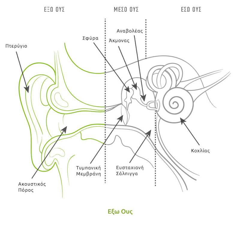
Μέσο Ους (Τύμπανο)
Ο τυμπανικός υμένας είναι ένας κυκλικός δίσκος λεπτού δέρματος με διάμετρο 8-9mm, κωνικού σχήματος με την κορυφή προς τα μέσα. Το μέσο ους αυτό καθαυτό βρίσκεται προς το έσω του τυμπανικού υμένα και είναι ένας αεροβριθής χώρος που περιέχει τρία μικρά οστά (σφύρα, άκμονας και αναβολέας), τα οποία συνδέουν τον τυμπανικό υμένα με το έσω ους.
Η σφύρα έχει μια λαβή και μια κεφαλή και η λαβή είναι τοποθετημένη ανάμεσα στις στιβάδες του τυμπανικού υμένα. Η κεφαλή της σφύρας εφάπτεται σε ένα χώρο του μέσου ωτός που ονομάζεται επιτυμπάνιος χώρος και αρθρώνεται στο μάλλον ογκώδες σώμα του άκμονα. Από τον άκμονα ξεκινά μια αποφυάδα που στρέφεται προς το κάτω τοίχωμα του μέσου ωτός και τελικά συνδέεται με την κεφαλή του αναβολέα. Τα δύο σκέλη του αναβολέα συνδέονται στη βάση του, η οποία βρίσκεται μέσα σε μια μικρή οπή, την ωοειδή θυρίδα. Ακριβώς κάτω από την ωοειδή θυρίδα υπάρχει και μια άλλη μικρή οπή προς το έσω ους, η οποία ονομάζεται στρογγυλή θυρίδα. Ένας λεπτός υμένας κλείνει την οπή αυτή και όταν η βάση του αναβολέα κινείται «μέσα-έξω» ο υμένας της στρογγυλής θυρίδας κινείται «έξω-μέσα» επειδή το υγρό στο έσω ους μεταδίδει τις μεταβολές της πίεσης.
Διαμέσου του μέσου ωτός πορεύεται το προσωπικό νεύρο, το οποίο εγκαταλείπει τον εγκέφαλο και περνάει από το κρανίο ώστε να νευρώσει τους μύες της έκφρασης του προσώπου.
Στο μέσο ους υπάρχουν και δύο μικροί μύες. Ο ένας από αυτούς βρίσκεται στο πρόσθιο τμήμα, ο τείνων το τύμπανο μυς, του οποίου η λειτουργία δεν είναι σαφής, ενδέχεται να στοχεύει απλώς να κάνει τη σίτιση και την κατάποση λιγότερο θορυβώδης. Ο δεύτερος, ο μυς του αναβολέα, εντοπίζεται στο οπίσθιο τμήμα του μέσου ωτός και αντιδρά σε δυνατούς ήχους με σύσπαση και σύσφιξη της αλυσίδας των ακουστικών οσταρίων και πιθανώς, μειώνει τη μετάδοση παρατεταμένων και δυνητικά βλαβερών ήχων προς το έσω ους.
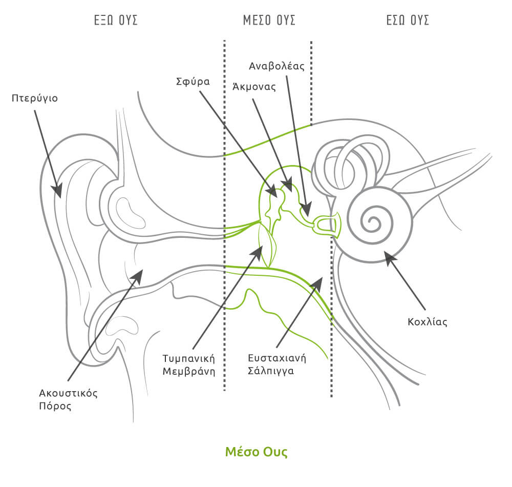
Έσω ους
Το έσω ους είναι, ίσως, το πιο πολύπλοκο μέρος του ανθρώπινου σώματος. Κάνει δυνατή την ακοή μετατρέποντας τον ήχο σε ηλεκτρικά σήματα, τα οποία στη συνέχεια ταξιδεύουν κατά μήκος του ακουστικού νεύρου στον εγκέφαλο. Επιπλέον, το έσω ους παίζει ρόλο και στην ισορροπία μας, καθώς χάρη στο λαβύρινθο του αυτιού μπορούμε να αντιληφθούμε την επιτάχυνση του κεφαλιού σε κάθε διεύθυνση, είτε σε ευθεία γραμμή είτε σε περιστροφή ή στροφή.
Το τμήμα του έσω ωτός που πραγματικά «ακούει» είναι ο κοχλίας. Ο σωλήνας αυτός είναι γεμάτος με ένα υγρό, επονομαζόμενο ως έξω λέμφος, όμοιο με αυτό που κυκλοφορεί σε ολόκληρο το σώμα και το υγρό που περιβάλλει τον εγκέφαλο. Μέσα στην έξω λέμφο βρίσκεται ο κοχλιακός πόρος, τα τριχωτά κύτταρα και σε επαφή με τη βάση αυτών είναι οι νευρικές ίνες που μεταφέρουν τις ώσεις (σήματα) προς τον εγκέφαλο.
Περίπου οι μισές από τις ακουστικές ίνες περνούν στην αντίθετη πλευρά του εγκεφαλικού στελέχους και στη συνέχεια περνούν στον μεσεγκέφαλο, για να φτάσουν τελικά οι πληροφορίες που μεταφέρουν στον φλοιό των εγκεφαλικών ημισφαιρίων, όπου γίνονται συνειδητές.